Pituitary tumors are among the most commonly diagnosed tumors affecting the base of the brain. Although many pituitary tumors are noncancerous, they can cause significant health problems when they grow, produce excessive hormones, or press against nearby structures such as the optic nerves. Depending on the tumor’s size, location, and characteristics, surgery may be recommended to remove the tumor and protect surrounding structures.
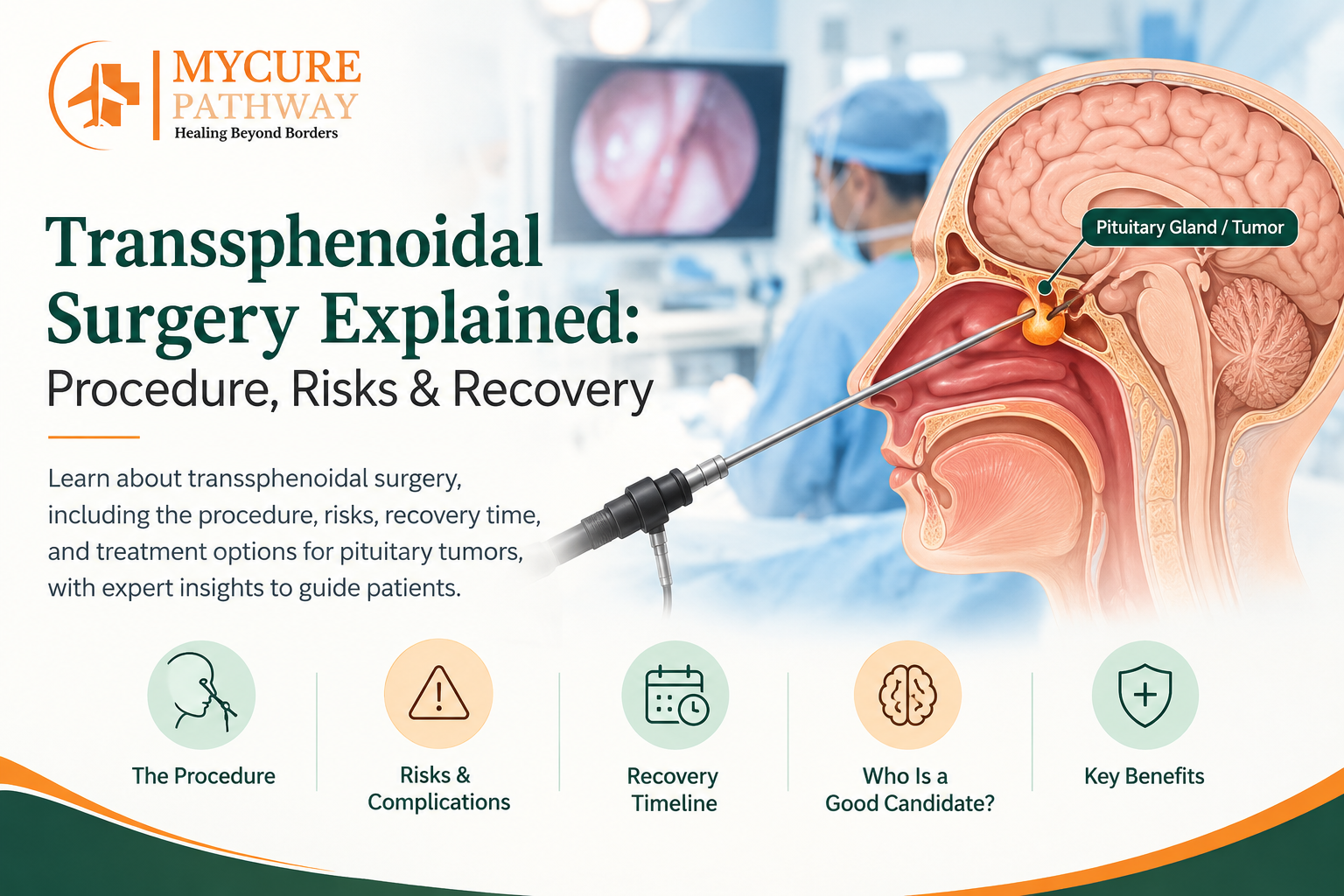
Transsphenoidal surgery is a commonly used surgical approach for treating selected pituitary tumors. Instead of opening the skull, surgeons reach the pituitary gland through the nasal passages and sphenoid sinus. Advances in surgical technology have introduced two important techniques: microscopic transsphenoidal surgery and endoscopic transsphenoidal surgery.
Both techniques aim to remove pituitary tumors while minimizing damage to surrounding tissues. However, they differ in visualization, surgical instruments, operating techniques, and the way surgeons access difficult-to-reach areas.
For patients exploring endoscopic transsphenoidal surgery, understanding how it compares with the traditional microscopic approach can help them make informed treatment decisions. This article explains the major differences, benefits, limitations, recovery expectations, and factors that influence surgical outcomes.
What Is Transsphenoidal Surgery?
Transsphenoidal surgery is a specialized neurosurgical procedure used to access the pituitary gland and other selected abnormalities near the skull base.
The pituitary gland is a small, pea-sized structure located beneath the brain. It produces hormones that regulate several important bodily functions, including growth, metabolism, reproduction, thyroid activity, and the body’s response to stress.
The term transsphenoidal refers to the surgical route through the sphenoid sinus, an air-filled cavity located behind the nose.
This approach is frequently used to treat pituitary adenomas, which are usually noncancerous growths arising from the pituitary gland. Depending on the patient’s condition, surgery may also be considered for selected Rathke’s cleft cysts, craniopharyngiomas, and other lesions around the sellar region.
The main objectives of transsphenoidal surgery include:
- Removing as much of the tumor as safely possible.
- Relieving pressure on the optic nerves and nearby structures.
- Reducing excessive hormone production from functioning tumors.
- Preserving healthy pituitary tissue whenever possible.
- Improving symptoms and preventing further complications.
Unlike conventional open brain surgery, the transsphenoidal approach does not usually require a large opening in the skull. However, it remains a complex neurosurgical procedure that requires specialized training and careful postoperative monitoring.
What Is Endoscopic Transsphenoidal Surgery?
Endoscopic transsphenoidal surgery is a minimally invasive pituitary surgery technique that uses a thin endoscope equipped with a camera and light source to access the pituitary gland through the nose.
The endoscope transmits magnified images to a monitor, allowing the surgeon to examine the surgical area and use specialized instruments to remove the tumor.
The procedure is commonly performed through one or both nostrils, depending on the surgical plan and the tumor’s anatomy.
How Does the Endoscopic Endonasal Approach Work?
The endoscopic endonasal approach allows surgeons to reach the pituitary gland through the natural nasal passages without creating a conventional opening in the skull.
The procedure generally involves the following steps:
1. General Anesthesia
The patient receives general anesthesia and remains unconscious throughout the operation. The surgical team monitors vital signs and prepares the nasal passages.
2. Introducing the Endoscope
The surgeon carefully inserts a thin endoscope through the nostril. The camera provides magnified images of the nasal passages and surrounding structures.
3. Accessing the Sphenoid Sinus
The surgeon navigates through the nasal corridor and opens the sphenoid sinus to reach the area containing the pituitary gland.
4. Accessing the Pituitary Tumor
A small opening is created in the bone covering the pituitary gland. Specialized surgical instruments are then used to access the tumor.
5. Tumor Removal
The surgeon removes the tumor under continuous endoscopic visualization. The aim is to achieve the maximum safe removal while protecting important structures, including the optic nerves, normal pituitary tissue, and nearby blood vessels.
6. Surgical Reconstruction
After tumor removal, the surgeon checks for bleeding and cerebrospinal fluid leakage. If necessary, the surgical opening is repaired using tissue grafts or other reconstruction techniques.
7. Postoperative Monitoring
The patient is transferred to the recovery area for monitoring. Doctors assess neurological function, hormone levels, fluid balance, and possible complications.
Key Advantages of Endoscopic Transsphenoidal Surgery
Endoscopic surgery offers several potential advantages:
- Wide-angle visualization: The endoscope provides a broad view of the surgical field.
- Improved visualization of certain areas: Angled endoscopes can help surgeons examine regions that may be difficult to see through a straight microscopic view.
- Magnified images: High-definition cameras provide detailed visualization of anatomical structures.
- No large external incision: The procedure uses the nasal passages to access the tumor.
- Potentially less tissue disruption: The endonasal approach can reduce disruption of some surrounding tissues compared with traditional microscopic techniques.
- Collaborative surgical approach: Neurosurgeons and ENT surgeons may work together to manage different stages of the procedure.
However, the advantages depend on the patient’s anatomy, tumor characteristics, surgical technique, and the experience of the operating team.
What Is Microscopic Transsphenoidal Surgery?
Microscopic transsphenoidal surgery is a traditional technique used to remove pituitary tumors through the nasal passage and sphenoid sinus.
Instead of using an endoscope and camera, the surgeon operates under a surgical microscope that provides magnified, illuminated visualization of the surgical area.
This technique has been used for decades and remains an important option in selected patients.
How Is Microscopic Transsphenoidal Surgery Performed?
The general procedure follows a similar route to endoscopic surgery, but the visualization system and instruments differ.
1. Anesthesia and Preparation
The patient receives general anesthesia. The surgeon prepares the nasal passage and establishes the surgical corridor.
2. Access Through the Nasal Passage
A nasal incision may be made, depending on the specific microscopic technique. Specialized instruments are used to reach the sphenoid sinus.
3. Microscopic Visualization
The surgeon uses an operating microscope to obtain a magnified view of the surgical area.
4. Opening the Sellar Region
A small opening is made in the bone surrounding the pituitary gland to access the tumor.
5. Tumor Removal
Using specialized instruments, the surgeon removes the tumor while observing the operative area through the microscope.
6. Closure and Recovery
After tumor removal, the surgeon checks the area for bleeding or cerebrospinal fluid leakage and performs the necessary reconstruction before completing the procedure.
Advantages of Microscopic Transsphenoidal Surgery
Microscopic surgery has several established advantages:
- It is a well-established surgical technique with a long history of clinical use.
- Surgeons familiar with the approach may benefit from extensive experience.
- The microscope provides magnified, three-dimensional depth perception through binocular viewing.
- It can be effective for appropriately selected pituitary tumors.
- It may be suitable when the tumor’s location and anatomy allow safe access through the microscopic corridor.
However, the microscope provides a relatively fixed line of sight. Some areas outside the direct viewing angle may be difficult to examine without repositioning instruments or adjusting the surgical approach.
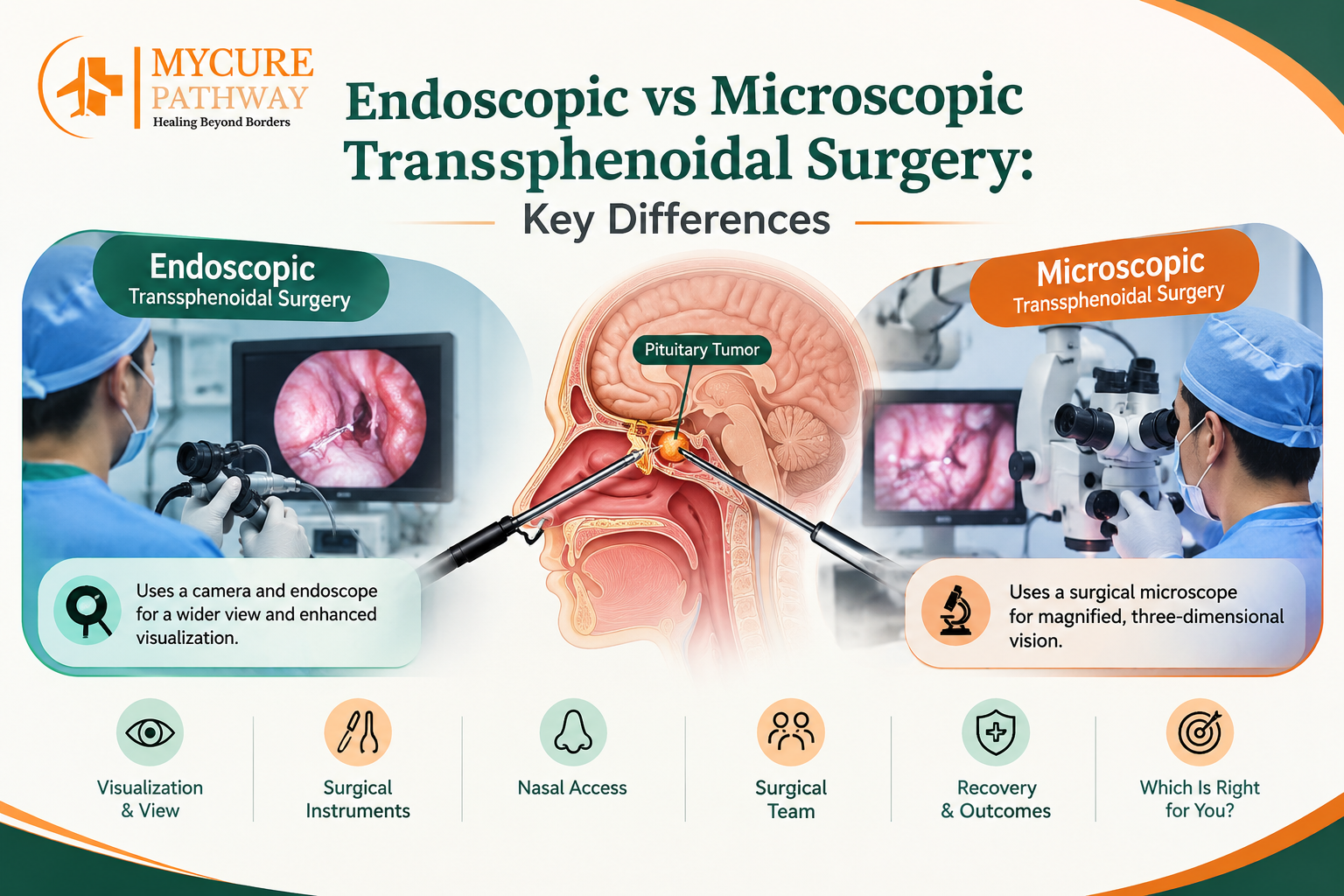
Endoscopic vs Microscopic Transsphenoidal Surgery: Key Differences
Although both procedures use the nasal route to reach the pituitary gland, they differ in important ways.
The choice depends on the tumor’s location, size, extension, the surgeon’s experience, and available technology.
| Comparison factor | Endoscopic surgery | Microscopic surgery |
| Visualization | Camera-based endoscopic images | Operating microscope |
| Field of view | Wide-angle view | Relatively narrow, direct view |
| Surgical instruments | Long, specialized endoscopic instruments | Instruments designed for microscopic access |
| Angled visualization | Angled endoscopes can examine hidden areas | More dependent on the microscope’s viewing axis |
| Nasal access | Often uses one or both nostrils | Traditionally uses a more restricted surgical corridor |
| Surgical collaboration | Frequently involves ENT and neurosurgical teams | May be performed by a neurosurgeon, depending on the technique |
| Depth perception | Requires interpretation of camera-based images | Binocular microscopic visualization |
| Tumor suitability | Suitable for a wide range of selected sellar and skull-base lesions | Effective for selected pituitary tumors |
| Recovery | Depends on surgical extent and complications | Depends on surgical extent and complications |
Neither technique is automatically the best choice for every patient. Both require careful surgical planning and experienced specialists.
Which Technique Provides Better Visualization?
Visualization is one of the most important differences between endoscopic and microscopic transsphenoidal surgery.
The endoscope provides a camera-based view of the surgical field. Its wide-angle perspective and availability of angled lenses can help surgeons examine areas beyond the direct line of sight.
This can be particularly useful when tumors extend toward the cavernous sinus or other surrounding regions.
The microscope, on the other hand, provides binocular magnification and a direct view of the surgical area. This offers natural depth perception, which some surgeons find valuable when working with delicate structures.
The main limitation is that the microscope’s viewing angle can restrict visualization of certain areas.
With endoscopic surgery, the surgeon may need additional training to interpret two-dimensional monitor images and understand depth and spatial relationships.
Ultimately, the quality of visualization depends on the equipment, surgical technique, anatomical conditions, and the surgeon’s experience.
Endoscopic vs Microscopic Surgery: Which Is Better for Pituitary Tumors?
There is no single answer because the most appropriate approach depends on the tumor’s characteristics.
Small Pituitary Tumors
Both techniques can be effective for appropriately located small pituitary adenomas.
The surgeon considers the tumor’s position, hormone production, and relationship with normal pituitary tissue when choosing the surgical method.
Large Pituitary Tumors
Larger pituitary tumors may extend beyond the normal boundaries of the pituitary gland and affect nearby structures.
Endoscopic surgery can provide broader visualization, which may be helpful when dealing with complex tumor extensions.
However, some large or invasive tumors may not be completely removable through either approach.
Tumors Extending Toward the Cavernous Sinus
The cavernous sinus contains important blood vessels and nerves. Tumors extending into this region can be particularly challenging to remove safely.
Angled endoscopes may help surgeons examine portions of this area. Nevertheless, the presence of critical structures can limit the extent of safe tumor removal.
Hormone-Producing Pituitary Tumors
Some pituitary adenomas produce excessive hormones, leading to conditions such as acromegaly or Cushing disease.
The primary goal of surgery is to remove the hormone-producing tumor and achieve biochemical control while preserving normal pituitary function.
The success of treatment depends on tumor biology, size, location, and the completeness of removal. The surgical approach alone does not determine the outcome.
Minimally Invasive Pituitary Surgery: Is Endoscopic Surgery Less Risky?
Endoscopic transsphenoidal surgery is often described as minimally invasive because it accesses the pituitary gland through the nasal passages without requiring a conventional opening in the skull.
However, minimally invasive does not mean risk-free.
Both endoscopic and microscopic procedures involve operating near important structures, including the brain, optic nerves, pituitary gland, and major blood vessels.
Potential complications include:
1. Cerebrospinal Fluid Leak
Cerebrospinal fluid (CSF) surrounds and protects the brain and spinal cord. A leak may occur if the protective tissue around the brain is opened during surgery.
Some leaks require additional treatment or surgical repair. Persistent leakage can increase the risk of meningitis.
2. Hormonal Imbalances
The pituitary gland regulates several hormones. Surgery may temporarily or permanently affect its ability to produce them.
Some patients require long-term hormone replacement therapy.
3. Diabetes Insipidus
Damage or temporary disturbance of the hormone-regulating system can cause arginine vasopressin deficiency, traditionally called central diabetes insipidus.
Symptoms may include excessive urination, increased thirst, dehydration, and abnormal sodium levels.
4. Vision-Related Complications
Because the optic nerves are close to the pituitary gland, injury to these structures may cause visual disturbances.
In many patients, surgery is intended to relieve pressure on the optic nerves, but new or worsening visual problems are possible.
5. Bleeding and Infection
Bleeding may occur during or after surgery. Infection is another potential complication, although serious infections are uncommon.
6. Nasal Complications
Patients may experience nasal congestion, crusting, reduced sense of smell, or discomfort during recovery.
7. Residual or Recurrent Tumor
Complete tumor removal may not be possible, especially when the tumor extends into surrounding structures. Some tumors may also recur after apparently successful surgery.
Regular imaging and hormone assessments are important for long-term monitoring.
The overall risk profile depends on the patient’s health, tumor characteristics, surgical complexity, and the experience of the surgical team.
Transsphenoidal Surgery Success Rate: What Determines the Outcome?
The transsphenoidal surgery success rate varies depending on the type of pituitary tumor, its size, hormone activity, location, and whether complete removal is possible.
It is important to understand that surgical success can mean different things:
- Complete or near-complete tumor removal.
- Improvement in vision or neurological symptoms.
- Normalization of excessive hormone production.
- Preservation of normal pituitary function.
- Long-term control without additional treatment.
For example, a small, non-invasive pituitary adenoma may be easier to remove completely than a large tumor extending into the cavernous sinus.
Similarly, hormone-producing tumors may require biochemical remission as an important measure of success, rather than relying only on postoperative MRI findings.
Does Endoscopic Surgery Have a Higher Success Rate?
Some studies have reported improved visualization and favourable outcomes with endoscopic techniques, particularly for selected complex pituitary and skull-base tumors.
However, it is not accurate to assume that endoscopic surgery always has a higher success rate than microscopic surgery.
Comparisons between the two techniques can be affected by:
- Differences in tumor size and complexity.
- The experience of the operating surgeon.
- Improvements in surgical technology over time.
- Differences in patient selection.
- The definition of surgical success used in individual studies.
- The duration of postoperative follow-up.
Patients should ask their surgeon about outcomes for tumors similar to their own, including the likelihood of complete removal, hormone remission, complications, and the possibility of additional treatment.
Recovery After Endoscopic and Microscopic Transsphenoidal Surgery
Recovery following both procedures is generally influenced more by the extent of surgery and the patient’s health than by the visualization method alone.
However, individual recovery experiences may differ.
First 24–48 Hours
Patients are monitored after surgery for neurological changes, bleeding, fluid balance, and hormonal disturbances.
Common symptoms include:
- Nasal congestion.
- Mild headaches.
- Fatigue.
- Nasal discomfort.
- Temporary changes in smell.
Doctors may perform blood tests to monitor sodium levels and pituitary hormone function.
First One to Two Weeks
Patients are generally advised to avoid activities that increase pressure inside the nasal passages.
These restrictions may include avoiding forceful nose blowing, heavy lifting, strenuous exercise, and straining during bowel movements.
Patients should follow their surgeon’s instructions regarding nasal cleaning, bathing, and medication.
Weeks Two to Six
Many patients gradually resume light activities, depending on their recovery.
Follow-up may include nasal examination, hormone testing, and assessment of symptoms. Some patients require additional treatment for hormone deficiencies or other complications.
Long-Term Follow-Up
MRI scans and hormone tests help doctors determine whether the tumor has been adequately treated and whether any residual growth requires monitoring.
Patients with functioning pituitary tumors may need continued endocrine assessment even after apparently successful surgery.
Seek urgent medical attention for persistent clear watery nasal drainage, severe or worsening headache, fever with neck stiffness, new vision changes, excessive thirst with frequent urination, or confusion.
How to Choose the Right Surgical Approach?
Choosing between endoscopic and microscopic transsphenoidal surgery requires a detailed evaluation by an experienced neurosurgeon.
Patients should consider several factors before proceeding.
1. Tumor Size and Location: The surgical approach should provide safe access to the tumor while protecting surrounding structures.
2. Tumor Extension: Tumors extending into nearby areas may require specialized techniques and additional surgical planning.
3. Hormone Activity: Functioning tumors may have different treatment goals compared with non-functioning adenomas.
4. Surgeon’s Experience: Experience with the proposed technique is an important consideration. Patients should ask about the surgeon’s experience with similar cases.
5. Hospital Facilities: Advanced imaging, neuroendocrine support, intensive care, and postoperative monitoring contribute to comprehensive treatment.
6. Long-Term Follow-Up: A good treatment plan should include hormone assessments, MRI monitoring, and access to additional treatment if required.
Patients should also understand that surgery is not necessary for every pituitary tumor. Some tumors can be monitored, while certain hormone-producing adenomas may respond to medication.
Why Consider Pituitary Tumor Treatment in India?
India has specialized neurosurgical centres offering treatment for pituitary tumors and other skull-base conditions.
Patients may access multidisciplinary care involving neurosurgeons, ENT surgeons, endocrinologists, radiologists, and neuro-ophthalmologists.
When comparing hospitals, patients should consider:
- Experience in endoscopic and microscopic pituitary surgery.
- Availability of advanced endoscopic equipment.
- Neurosurgical and ENT collaboration.
- Access to MRI and endocrine investigations.
- Postoperative intensive care and monitoring.
- Rehabilitation and long-term follow-up facilities.
International patients should share their MRI scans, hormone reports, previous treatment records, and medical history before travelling.
A personalized treatment recommendation and cost estimate should be obtained from the treating hospital. The final surgical approach must be determined by the specialist team after reviewing the patient’s clinical findings.
Frequently Asked Questions
1. Is endoscopic transsphenoidal surgery better than microscopic surgery?
Endoscopic surgery offers a wider field of view and can help surgeons examine areas that may be difficult to visualize with a microscope. However, both approaches can be effective for selected pituitary tumors. The best option depends on tumor anatomy and the surgeon’s expertise.
2. Is endoscopic transsphenoidal surgery considered minimally invasive?
Yes. It is generally considered a minimally invasive approach because the surgeon reaches the pituitary gland through the nasal passages rather than making a conventional opening in the skull. However, it remains a major neurosurgical procedure with potential complications.
3. How long does transsphenoidal surgery take?
Many procedures take approximately two to four hours, although complex cases may require additional time. The duration depends on tumor size, location, surgical complexity, and reconstruction requirements.
4. What is the success rate of endoscopic transsphenoidal surgery?
Success rates vary according to tumor type, size, location, hormone activity, and surgical expertise. Small, non-invasive tumors generally have more favourable removal outcomes than large or invasive tumors. Patients should discuss case-specific outcomes with their neurosurgeon.
5. Can pituitary tumors return after surgery?
Yes. Some pituitary tumors may recur or continue growing if a portion remains after surgery. Regular MRI scans and hormone testing help identify residual or recurrent disease.
Conclusion
Both endoscopic and microscopic transsphenoidal surgery are established approaches for treating selected pituitary tumors. While microscopic surgery uses a magnified direct view, endoscopic transsphenoidal surgery uses a camera-based system that provides a wider field of visualization and access to certain difficult-to-reach areas.
The endoscopic endonasal approach has become an important option in modern pituitary surgery because of its visualization capabilities and minimally invasive access. However, the most appropriate technique depends on the patient’s tumor characteristics, medical condition, and the experience of the surgical team.
Patients considering minimally invasive pituitary surgery should discuss the expected benefits, potential risks, transsphenoidal surgery success rate, and long-term follow-up requirements with a qualified neurosurgeon. A personalized treatment plan can help ensure that the chosen approach prioritizes safe tumor removal and preservation of neurological and hormonal function.
Explore More: Can a Brain Tumor Be Cured Without Surgery? What Specialists Say